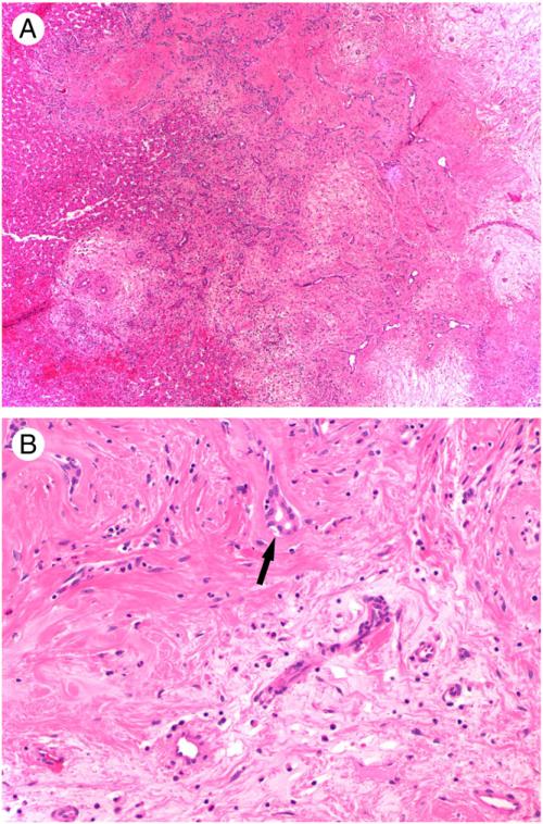
Fig. 2.
Histological features of MH. A, Nodular collection of hypocellular myxoid and hyalinized stroma with proliferating bile ducts (original magnification, ×40). Note the presence of normal-appearing hepatocytes at the periphery. B, Higher power view of myxoid (bottom) and hyalinized (upper) stroma with compressed bile ducts (black arrow), sparse bland spindle cells, and admixed inflammatory cells (original magnification, ×200).

