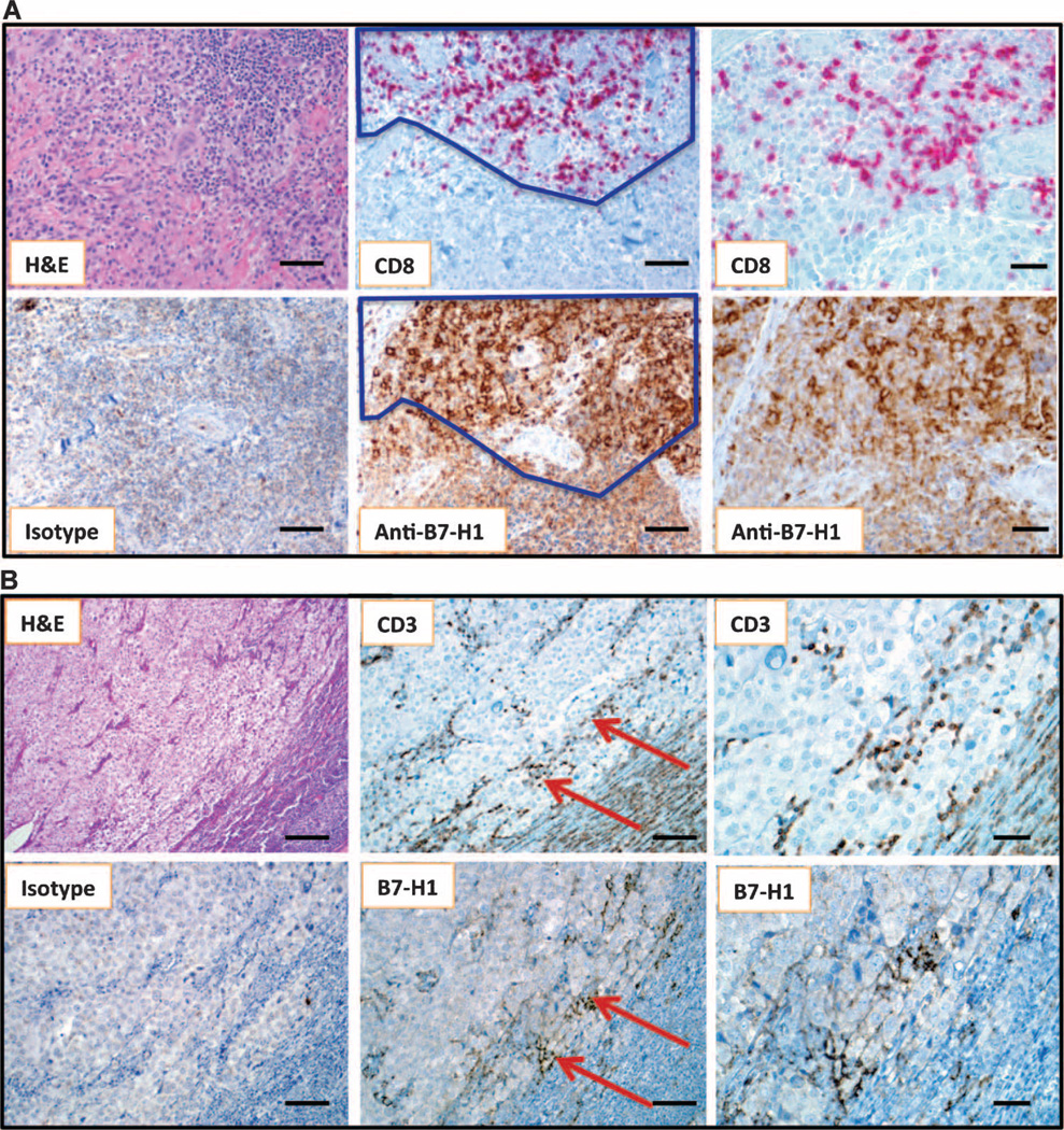
Fig. 2.
Geographic association of B7-H1 expression and TILs. (A) Primary nodular melanoma with associated “severe” grade of lymphocytic infiltration, highlighted by CD8 immunostaining. The dark line demarcates the area of colocalizing T cells and B7-H1+ tumor cells. Overall, 30% of the tumor cells demonstrated cell surface B7-H1 expression. Note that the melanoma cell cytoplasm is heavily pigmented due to abundant melanin (see isotype control). Original magnifications, ×200 for left and middle column panels (scale bars, 50 µm) and ×400 for right column panels (scale bars, 25 µm). (B) Metastatic deposit of melanoma in a lymph node, associated with “mild” grade of lymphocyte infiltration shown by CD3 immunostaining. Original magnifications, ×100 for left column panels (scale bars, 100 µm), ×200 for middle column panels (scale bars, 50 µm), and ×400 for right column panels (scale bars, 25 µm). Arrows highlight the geographic area of concordance of CD3+ TILs with B7-H1 expression by melanoma, and also point to the area shown at higher magnification in the right-hand panels. Five percent of tumor cells demonstrated B7-H1 expression in this case.

