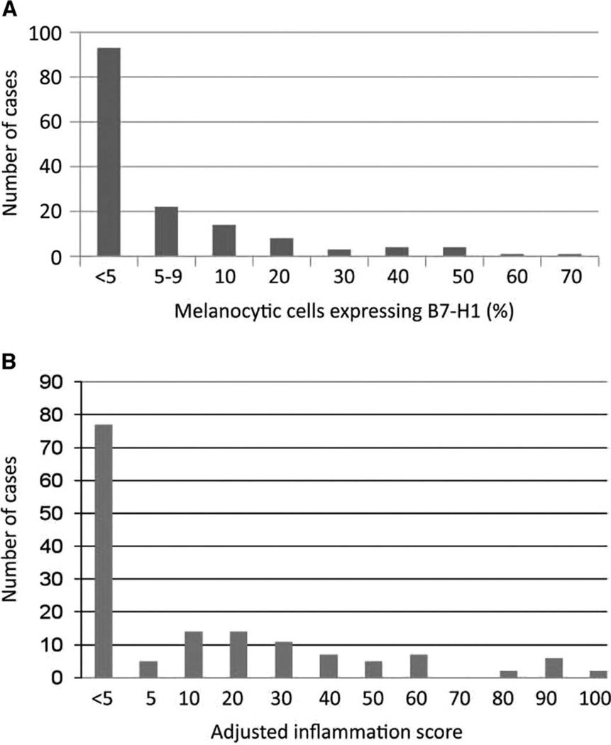
Fig. 3.
Distribution of intensity of B7-H1 expression by melanocytic cells and immune infiltrates in 150 lesions. (A) Percent B7-H1 expression by melanocytes ≥5% was considered positive. (B) AIS, defined as the intensity of intratumoral inflammation including TILs and histiocytes (graded 0 to 3, see text) multiplied by the percent B7-H1+ inflammatory cells.

