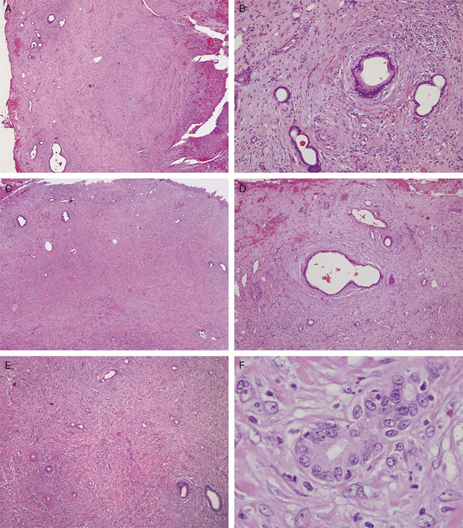
FIGURE 1.
Low-power view of the gallbladder (A) shows mucosal ulceration (right) and lobular aggregates of Luschka ducts in the subserosal soft tissue. At intermediate power (B), one can appreciate the bland cytology and concentric periductal fibrosis. Low-power view of another area in the gallbladder subserosa (C) shows usual Luschka ducts (left) merging with inflamed Luschka ducts showing florid reactive atypia (right). The more typical uninflamed Luschka ducts (D) maintain a lobular architecture and bland cytology. The inflamed Luschka ducts appear more disorganized in the inflamed nonconcentrically fibrotic stroma (E) and demonstrate significant cytologic atypia (F), best considered reactive.

