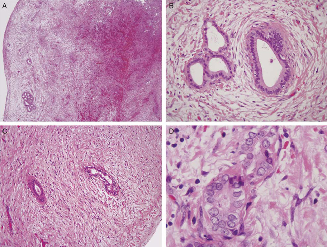
FIGURE 2.
At low power, the Luschka ducts appear as lobular aggregates of ducts within the gallbladder subserosa (A). At higher power magnification (B), the glands are surrounded by concentric fibrosis, maintain a lobular architecture, and demonstrate bland cytology. Individual fields of this case demonstrated distorting fibrosis (C) and mild cytologic atypia (D), which raised the possibility of neoplasia.

