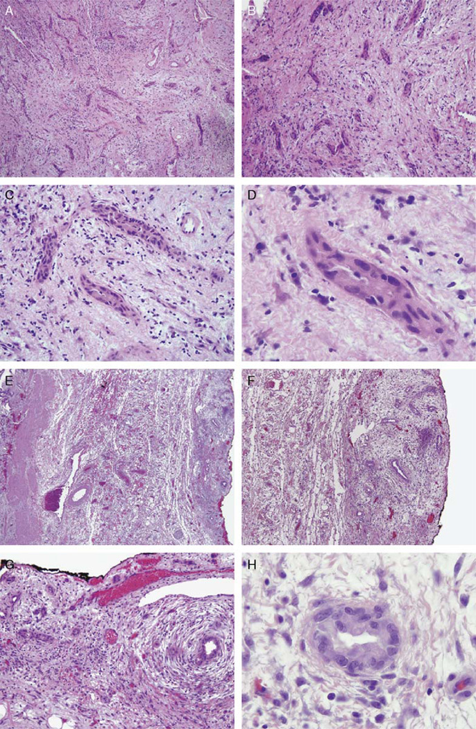
FIGURE 3.
The frozen section of the gallbladder fossa for case 1 shows a somewhat irregular proliferation of glands (A,B) with significant cytologic atypia (C,D) in a reactive stroma lacking concentric fibrosis, which suggested the diagnosis of metastatic adenocarcinoma involving the peritoneal cavity. The resected gallbladder specimen (E) contains similar glands now shown to be in a linear, lobular distribution along the gallbladder serosa (right), opposite the gallbladder lumen (left). These glands demonstrate concentric fibrosis (F) and show similar cytologic atypia (G,H). Given the stromal inflammation, these are consistent with as reactive Luschka ducts.

