FIGURE 4.
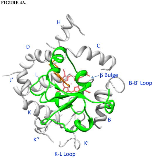
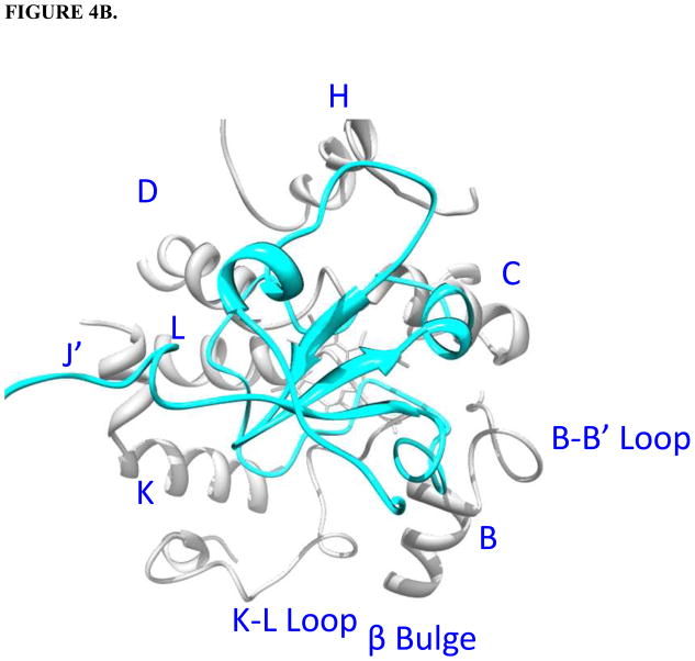
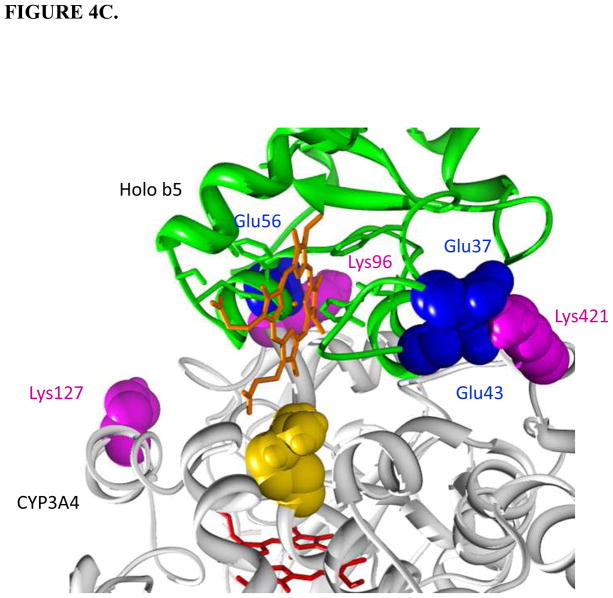
Holo/apo b5-CYP3A4 complex models. CYP3A4 is white; b5 is green; the heme group of CYP3A4 is red; the heme group of holo b5 is orange. The interacting residues on CYP3A4 and cyt b5 are magenta and blue, respectively. Protein regions on CYP3A4 far away from the interacting surfaces are truncated. A. Top view of holo b5-CYP3A4 model. B. Top view of apo b5-CYP3A4 model. C. R446A (golden) illustrated in the holo b5-CYP3A4 model.


