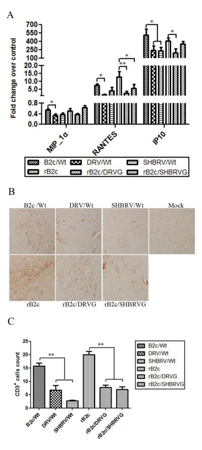
Fig. 4. Expression of chemokines and inflammatory responses in the CNS after infection with rRABV.
Groups of 3 female ICR mice at the age of 4 to 6 weeks were infected with each of the viruses at 10ICLD50 and transcardially perfused with 10% formalin at day 6 p.i. Brains were used for preparation of RNA and chemokine expression was assessed by qTR-PCR (A). Paraffin sections were subjected to immunohistochemistry for detecting CD3-positive cells (B). CD3-postive cells were quantified (C). Significance of differences between experimental groups was assessed by One-Way ANOVA. (*, p<0.05; **, p<0.01).

