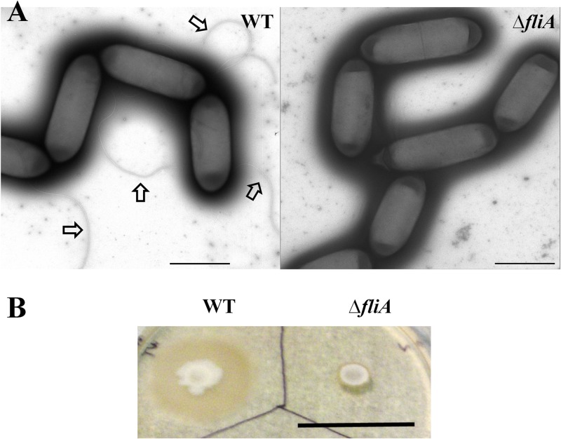
Fig 3.
Characterization of the fliA deletion mutant. (A) Production of flagella. Transmission electron micrographs of negatively stained C. ljungdahlii wild-type cells (left panel) and the fliA deletion mutant (right panel) showed that the mutant did not produce any flagella, whereas the wild-type cells produced flagella as indicated by arrows. Scale bars in both panels represent 2,000 nm. (B) Motility assay. The fliA deletion mutant was not motile when spotted on a YTF soft agar plate on which the wild-type cells were motile and thus exhibited a larger growth zone. The scale bar represents 3 cm.

