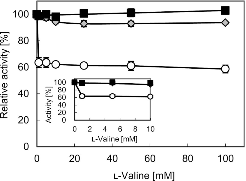
Fig 2.
Relative activity of the wild-type AHAS (IlvBN) and the mutant (IlvBNGM) in the presence of l-valine. Open circles, Val-4: IlvBN[plasmid]/IlvBN[chromosome]; gray diamonds, Val-5: IlvBNGE[plasmid]/IlvBN[chromosome]; black squares, Val-6: IlvBNGE[plasmid]/IlvBNGE[chromosome] (the location of ilvBN [wild type or mutant] on each strain is shown in the brackets). The inset shows the relative activity of AHAS in the presence of lower concentrations of l-valine. The data represent averages from three independent experiments.

