


Fig 7.
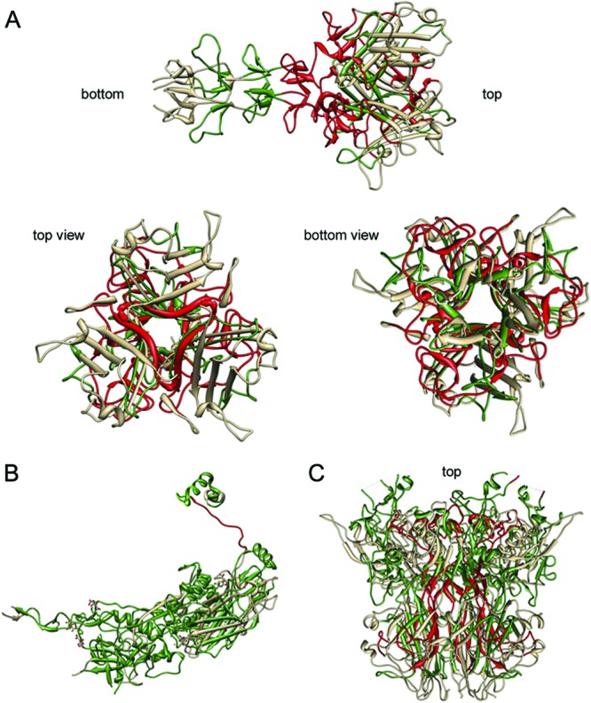
Visualizations of damaged peptides in protein structures of fiber (A), hexon (B), and penton (C) proteins after indirect sunlight disinfection (UVA+RB). Degraded peptides are shown in red, conserved peptides are shown in green, and peptides not covered by mass spectrometry are shown in gray. Images were created with Chimera (52).