Fig 2.

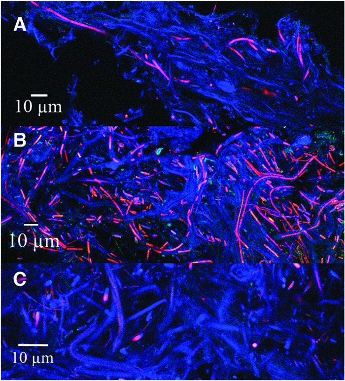
Confocal microscopy of coniform structures from Octopus Spring. (A) Tip of cone; (B) middle of cone; (C) bottom of cone. Chlorophyll a autofluorescence is shown in red; DAPI staining is shown in blue.

Confocal microscopy of coniform structures from Octopus Spring. (A) Tip of cone; (B) middle of cone; (C) bottom of cone. Chlorophyll a autofluorescence is shown in red; DAPI staining is shown in blue.