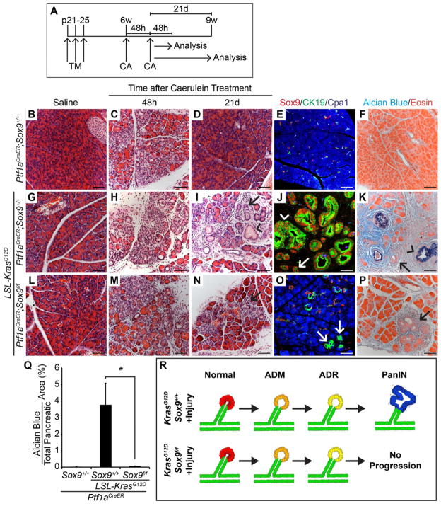
Figure 7. Sox9-deficient acinar cells expressing KrasG12D can undergo persistent acinar-to-ductal reprogramming (ADR) after acute pancreatitis, but do not progress into PanINs.
(A) Ptf1aCreER;Sox9+/+, Ptf1aCreER;LSL-KrasG12D;Sox9+/+ and Ptf1aCreER;LSL-KrasG12D;Sox9f/f mice were injected with tamoxifen (TM) at postnatal day (p) 21, 23 and 25. At 6 weeks (w) of age, mice were treated with two sets of caerulein (CA) or saline injections on alternating days and analyzed 48 hours (h) or 21 days (d) later. (B–D, G–I, L–N) H&E staining shows persistent acinar-to-ductal metaplasia (ADM) in Ptf1aCreER;LSL-KrasG12D;Sox9+/+ and to a lesser extent also in Ptf1aCreER;LSL-KrasG12D;Sox9f/f mice (I, N, arrows). PanINs are only present in Ptf1aCreER;LSL-KrasG12D;Sox9+/+ mice (I, arrowhead). (E, J, O) Co-immunofluorescence staining for CK19, Cpa1 and Sox9 reveals CK19+Cpa1− duct-like cell clusters in Ptf1aCreER;LSL-KrasG12D mice in the presence and absence of Sox9 (J, O, arrows). (F, K, P) Alcian blue and eosin staining shows PanINs in Ptf1aCreER;LSL-KrasG12D;Sox9+/+ mice (K), but not in Ptf1aCreER;LSL-KrasG12D;Sox9f/f mice (P). Arrows in K, P point to ADM and arrowheads in J, K to PanINs. (Q) Quantification of Alcian blue+ pancreatic area 21d after CA (n=4–5). (R) Schematic summarizing the phenotype observed in Sox9 loss-of-function experiments in the presence of KrasG12D and acute pancreatitis. Values are shown as mean ± s.e.m. *P<0.05. Scale bars: 50μm. See also Figure S5.

