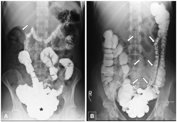
Figure 1. Imaging studies of the gastrointestinal tract.
A - Upper gastrointestinal series in upright standing position one hour after administration of oral contrast material. Note that the majority of contrast agent is within small bowel loops located deep in the pelvis, predominantly in the ileum (asterisk). Thus, despite upright position and contrast filling, the radiographic aspect suggests an abnormal downward displacement of small bowel into the pelvis. Notably, there is a large amount of stool in the right colon (arrow) causing a lack of contrast filling of the large bowel.
B - Barium enema in supine patient position after evacuation. Note an abnormal downward displacement and elongated appearance of the transverse colon into the pelvis even in supine position (arrows). The positioning of the ascending, descending and sigmoid colon appears to be normal. Due to evacuation the large bowel demonstrates variable distension and contrast pattern.

