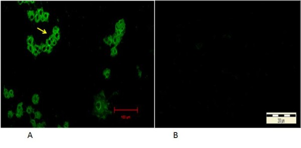
Figure 4.

Immunofluorescent antibodies staining of infected CrFK cells following infection with local FCoV UPM11C/08 isolate. The fluorescence signal appeared as granules covering areas within the cytoplasm at 24 hours PI (arrow). No signal is present in the nucleus. 100x Mag. Scale bar, 20μm (A). Absence of fluorescence signal in control uninfected cell cultures. 10x Mag. Bar = 200μm (B).
