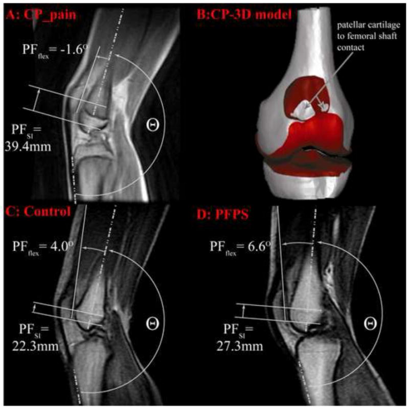
Figure 5. isual description of PF extension and patella alta at 20° of knee extension.

The knee angle was defined at 180°− Θ. The top row represents at single participant with CP and anterior knee pain. This participant had the most severe alta of the entire CP cohort. Image A: the cine-phase contrast (PC) magnetic resonance (MR) anatomic image. The patellofemoral superior displacement and extension is visually approximated on the image. Patellar extension can be approximated by its 2D counterpart (the angle between the vector bisecting the anterior and posterior edges of the distal femur and the vector delineating the posterior edge of the patella). The patellar superior position is the superior displacement of the patellar origin relative to the femoral origin (Figure 2) in the femoral coordinate system. Image B: A 3D model of the knee joint for this same individual at 20° knee extension. The patella is not shown, so that the contact between the patellar cartilage and femoral shaft can be seen. Image C: Cine-PC MR image for a single control subject with the 3rd highest level of patellar superior position out of all the control subjects. Image D: A cine-PC MR image from an otherwise healthy patient with PFPS from a previous study.24 Out of all the patients with PFPS in this previous study, this subject had the second highest level of patella alta.
