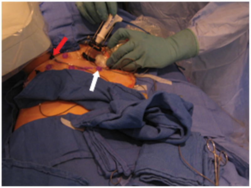
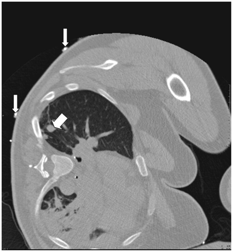
Fig. 1.


A. Close-up view of the tracking apparatus. Passive fiducials (red arrow) and active fiducials (white arrow) are located within the acquisition area of the field generation (top left hand corner of the figure). These fiducials are used for registration of the tracking system with pre-procedural CT images. B. Planning CT scan illustrates a solitary 12 mm melanoma metastasis in the right lung (thick arrow). Fiducials (thin arrows) are used in the registration process.
