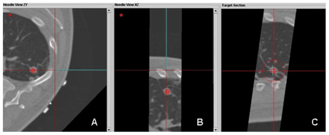
Fig. 3.

Custom tracking software displaying multiplanar views during tracked guider needle placement; operator is now viewing (not blinded to) the real-time tracking display. A. An axial plane shows the virtual needle (blue line) has a straight trajectory to the lesion. B. Para-coronal plan in the in the same plane as the needle, illustrates that the lesion and the needle path are in the same plane. C. “View down the needle shaft” shows that the needle tip (blue “+”) is directly in line with the target lesion (red dot).
