
FIG. 1.
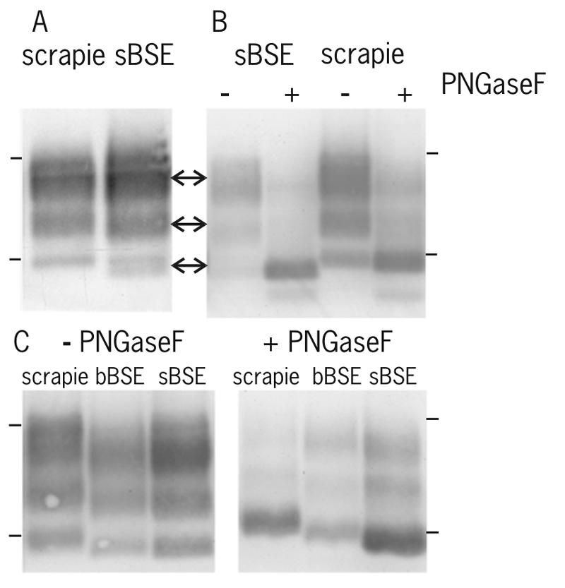
Migration patterns for PrPres in natural scrapie, experimental sheep BSE, and bovine BSE. (A) Sheep scrapie and sheep BSE (sBSE). The three glycosylation forms of PrPres, diglycosyl, monoglycosyl, and aglycosyl, are indicated by arrows, from top to bottom, respectively. (B) Comparison of migration positions for PrPres triplet bands before and after PNGaseF treatments. The positions of the aglycosyl band before (−) and after (+) deglycosylation by PNGaseF appear to be identical. An additional band below the aglycosyl form is variably present depending on the individual sample. (C) Differences in the migration of the PrPres triplet in sheep scrapie, bovine BSE (bBSE), and sBSE without (−) and with (+) PNGaseF treatments. Molecular mass markers were 29 and 18.4 kDa, and their migration positions are indicated by the upper and lower bars, respectively, beside the panels. The procedures used were the Prionics Check method (antibody 6H4) (A) and the methods designed for tissue treatment and Western blotting for this study (antibody 66.94b4) (B and C). Applied tissue equivalents were 430 μg (A) and 500 μg (with PNGaseF) and 1,000 μg (without PNGaseF) (B and C).