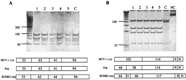
FIG. 1.
RFLP analysis of Argentine HCV isolates. Amplified DNA fragments were digested with HinfI and MvaI (A) or RsaI and HaeIII (B). (Top) Polyacrylamide gel electrophoresis. Lanes 1 to 5, untypeable isolates; lane C, Argentine isolate corresponding to genotype 1a/c; lane NC, negative control (251 bp). (Bottom) Restriction map of the isolates tested and prototypic HCV strains, as predicted from the DNA sequences (Webcutter program version 2.0). The numbers indicate the lengths of the restriction fragments (in base pairs). Genotypes are indicated in parentheses. HCV-1 (1a), prototypic HCV genotype 1 strain; Arg, untypeable Argentine isolates; EUHK2, prototypic HCV genotype 6 strain. The numbers on the left indicate the sizes of the standards (in base pairs).

