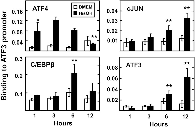
Fig. 6.
Chromatin immunoprecipitation (ChIP) analysis reveals temporal binding of regulatory factors at the ATF3 promoter during the AAR. HepG2 cells were maintained in DMEM ± 2 mM HisOH for 1, 3, 6, or 12 h, and then ATF4, cJUN, C/EBPβ, and ATF3 binding to the ATF3 proximal promoter was measured by ChIP analysis. The data are presented as the averages ± SD for a single experiment containing 3–4 replicates per condition, and each experiment was repeated with 2–3 independent batches of cells. *P value of ≤ 0.05 relative to the 1 h DMEM value, and **P value of ≤ 0.05 relative to the 1 h HisOH-treated value. Nonspecific IgG and primers specific for exon 2 of the ATF3 gene were used as negative controls and showed little or no binding (data not shown).

