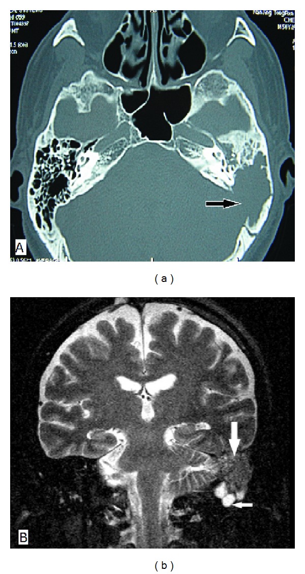
Figure 1.

Imaging findings of Case 1. (a) Axial CT of the temporal bone shows a soft-tissue mass in the left middle ear and mastoid cavity with extension into the sigmoid sinus plate (arrow); (b) coronal MRI shows a slightly low-signal-intensity mass on T2-weighted images in the left mastoid (large arrow). Several inferior lymph nodes are seen (small arrow).
