
Figure 2.
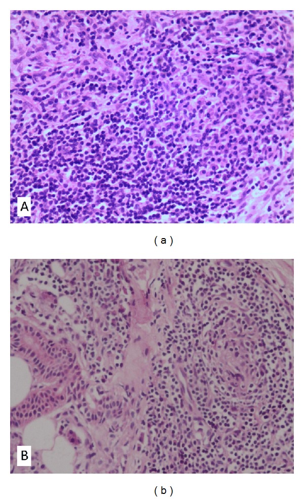
Histological pictures of Case 1. (a) Specimen obtained from the temporal bone demonstrates fibroblasts proliferation in an inflammatory infiltration background with predominance of plasma cells (H&E; original magnification ×200); (b) specimen of the parotid gland shows proliferation of lymphocytes, and plasma cells. A granuloma-like structure composed of fibroblasts, lymphocytes, and plasma cells was seen (H&E; original magnification ×200).