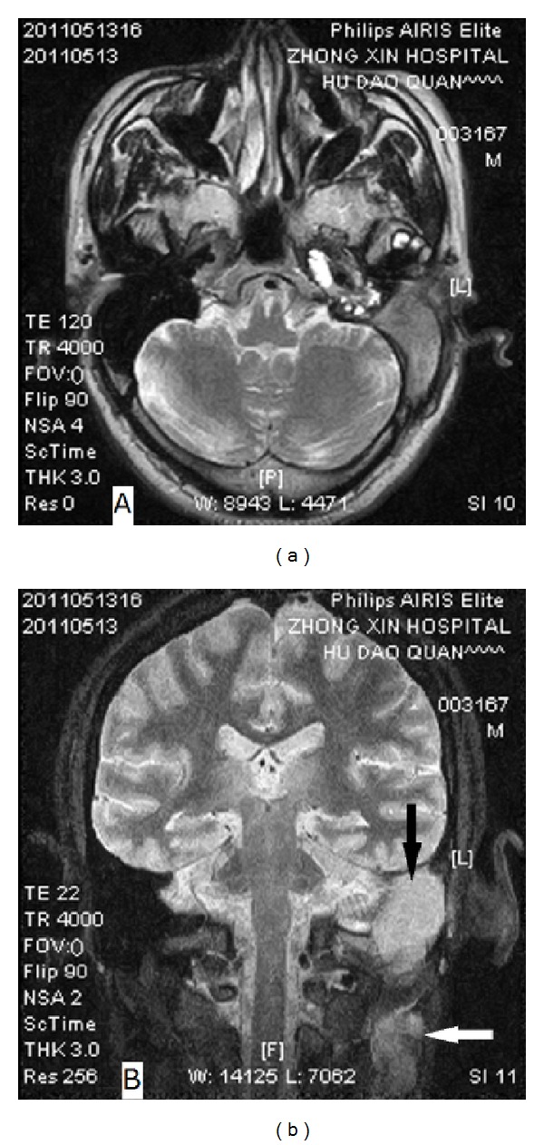
Figure 3.

MR images of Case 2. (a) T2-weighted axial image shows a hypointensity lesion in the left mastoid; (b) PDW coronal image demonstrates two nonhomogenous and slightly high-signal-intensity mass in the left mastoid (black arrow) and ipsilateral neck (white arrow), respectively.
