
Figure 2.
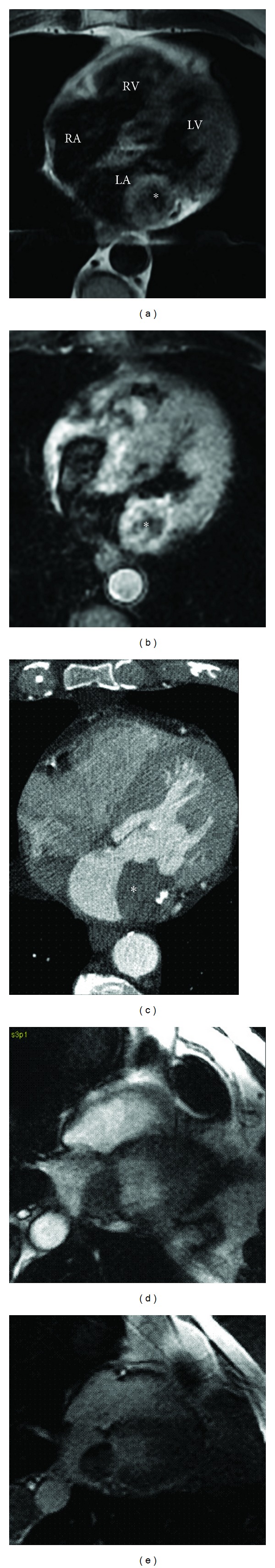
Axial (a) MRI T1- and (b) T2-weighted and (c) CT contrast enhanced imaging and 4-chamber MRI (d) first pass perfusion and (e) late gadolinium enhancement (TI = 350 ms) imaging. MRI T1- and T2-weighted black blood images demonstrate a lobulated mass (asterisk) with central signal drop-out suggestive of punctuate calcification confirmed by CT. CT imaging also suggests that this mass is contiguous with the posterior leaflet of the mitral valve. Dynamic contrast-enhanced MRI demonstrates very minimal first pass perfusion and heterogenous late enhancement consistent with surface thrombus overlying a tumour. (RA) right atrium, (LA) left atrium, (RV) right ventricle, and (LV) left ventricle.