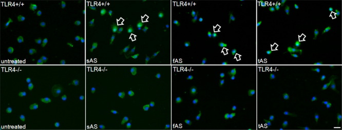
Figure 2.

NF-κB nuclear translocation upon AS treatment in TLR4+/+ microglia. Murine primary microglia were treated with different AS-forms (full length soluble AS–sAS, fibrillized AS–fAS, and C-terminally truncated AS–tAS). Cells were fixed with 4% paraformaldehyde, immunostained for NF-κB (green) and counterstained with DAPI (blue). NF-κB translocation in TLR4+/+ microglia treated with AS was identified by co-localization of DAPI and NF-κB (see arrows). No co-localization was observed in untreated and AS treated TLR4−/− microglia (scale bar, 10 μm).
