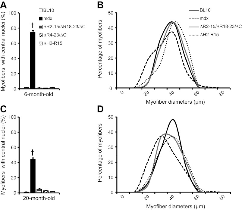
Fig. 3.
Quantitative characterization of extensor digitorum longus (EDL) muscle histopathology. A and B: 6 mo old. C and D: 20 mo old. The percentage of the myofiber with centrally localized nuclei in all 5 mouse strains is shown in A and C. Distribution of myofiber cross-sectional area in BL10, mdx, ΔR2-15/ΔR18-23/ΔC, and ΔH2-R15 is shown in B and D. †mdx mice are significantly different from all other age-matched mouse strains.

