
Fig. 3.
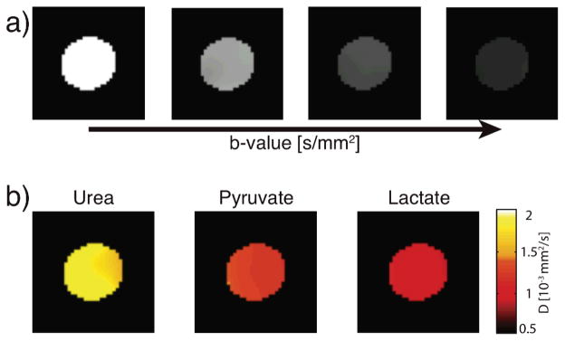
Diffusion weighted MRI of three hyperpolarized metabolites, acquired simultaneously. (a) Representative T1 corrected diffusion weighted images with increasing diffusion weighting (b-values 1.7, 173, 693 and 1560 s/mm2) used to simultaneously generate diffusion coefficient maps (b) of 13C urea, 13C pyruvate and 13C lactate.