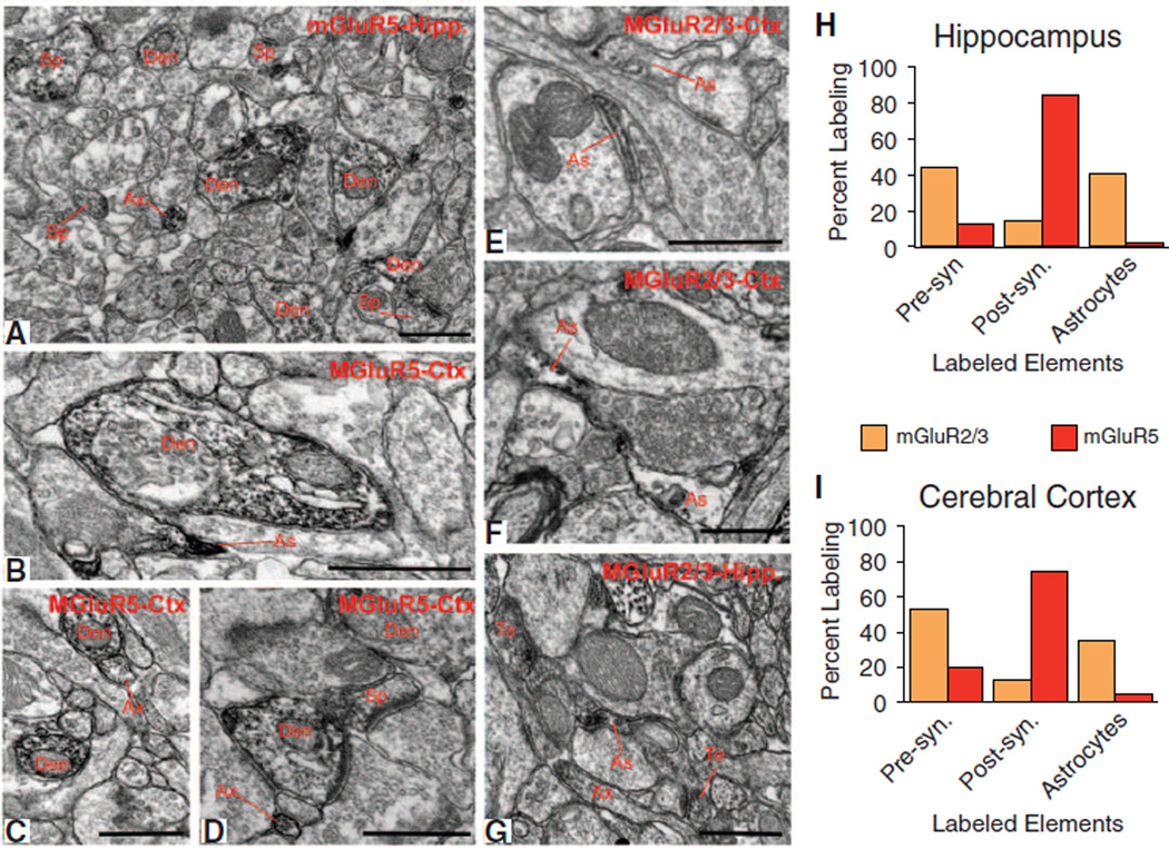
Fig. 2.
Electron micrographic analysis of mGluR2/3 and mGluR5 in the adult mouse cortex and hippocampus. (A to D) Electron micrographs showing examples of mGluR5-immunoreactive elements, and (E to G) mGluR2/3-immunoreactive elements in the mouse hippocampus (Hipp) and cerebral cortex (Ctx). Ax, unmyelinated axons; Den, dendrites; As, astrocytes; Te, axon terminals. Scale bars, 0.5 µm. (H and I) Histograms showing the relative percentage of mGluR5-or mGluR2/3-immunoreactive elements categorized as presynaptic (terminals and unmyelinated axons) or postsynaptic (dendrites and spines) neuronal elements or glia. For each region and antibody, data were collected in three mice. A total of 2215 µm2 of mGluR5 or mGluR2/3 immunostained tissue was examined in both cortex and hippocampus.

