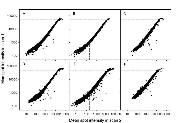
Figure 2.
Mean spot intensity in a scan with saturation (scan 1) versus mean spot intensities in a scan of the same arrays without saturation (scan 2), obtained at lower PMT voltage. ScanArray4000 I was used during scanning. (A), (B), and (C) Data of the red channel. (D), (E), and (F) Data of the green channel. (- - - - -) The intensity levels of 50 000 and 200. The PMT voltages (% of maximum voltage) used in scan 1 and scan 2 were 56% and 51% (A), 59% and 52% (B), 65% and 58% (C), 63% and 56% (D), 65% and 57% (E), 67% and 57% (F).

