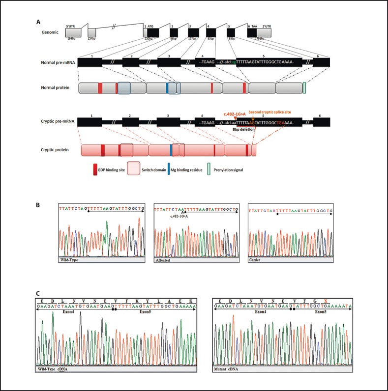
Fig. 3.
Schematic representation of RAB23 and molecular analysis of c.482-1G>A on splicing and protein translation. A Top: genomic organization of exons and introns of RAB23. Middle: wild-type pre-mRNA representation and functional domain in the 237 amino acid protein. Bottom: the truncated pre-mRNA and mutant protein. B Chromatograms of healthy control, homozygous and heterozygous individuals, respectively. Chromatograms show a novel substitution of G to A in the acceptor splice site of exon 5 (c.482-1G>A) depicted by a small arrow. The beginning of exon 5 is underlined. C Chromatograms of cDNA sequence analysis from patient (IV-2) and healthy control. The substitution alters the 3′ consensus splice site and activates a cryptic acceptor splice site downstream in exon 5 at position c.490. This results in the deletion of 8 nucleotides from the beginning of exon 5, causing a frameshift followed by a premature termination codon. X = Represents the premature termination codon. Nucleotide sequences of exon 4 and 5 are underlined.

