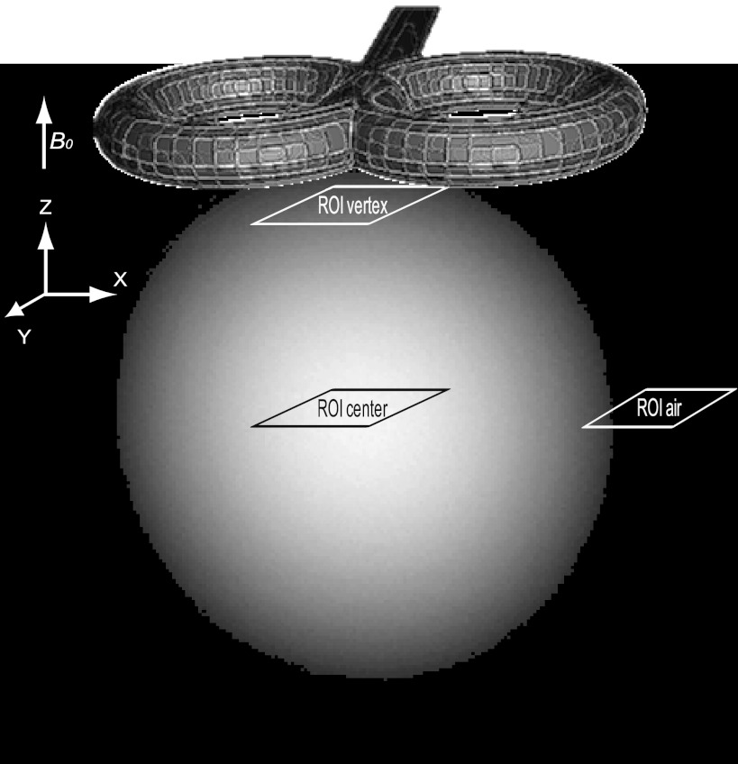
Fig. 1.
Position of transcranial magnetic stimulation (TMS) coil and regions of interest (ROIs). Anatomic image of the used phantom in which the TMS coil position is shown together with the ROIs used for computations of the signal-to-noise ratio (SNR) and for extraction of echo-planar imaging (EPI) time courses. B0, static magnetic field.

