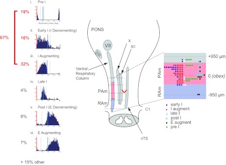
Fig. 7.

Summary of cell types and their spatial localization in PAm. Left: phase histograms of PAm respiratory-related neurons aligned vertically. Respiratory cycles are aligned to the onset of the inspiratory phase (red; 0°) to show the respective contribution of different cell types throughout the respiratory cycle. The time of occurrence of the IE transition (blue) could vary within the respiratory cycle. i, pre-I phase histogram; ii, early I phase histogram; iii, I-augment phase histogram; iv, late I phase histogram; v, post-I phase histogram; vi, E-augment phase histogram. Right: the rostrocaudal distribution of recorded respiratory-related cells with respect to the obex. The ventral respiratory column (VRC) and other brainstem nuclei are illustrated schematically in the brainstem drawing on the left. The rostrocaudal location and cell type are schematically represented in an enlargement of the portion of the VRC spanning the obex (red V) on the right. VII, facial nucleus; VIII, cochlear nucleus; X, dorsal motor nucleus of the vagus nerve; XII, hypoglossal nucleus; C1, spinal cord cervical section 1.
