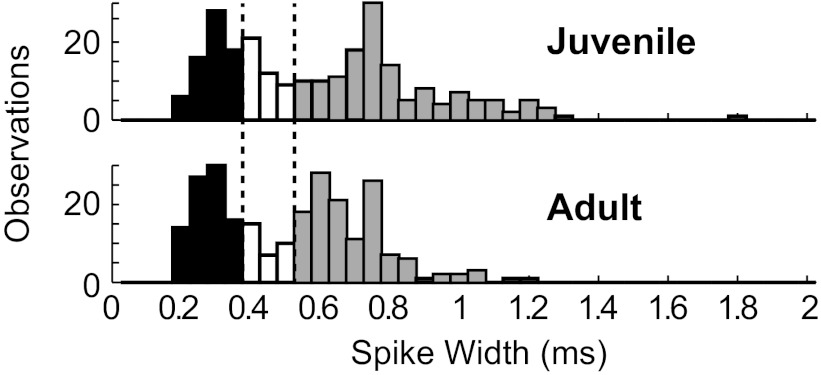
Fig. 2.
Histograms of HVC neuronal spike widths reveal a dip in the distribution between 0.35 and 0.50 ms (marked by dotted lines and open bars) in juveniles (top) and adults (bottom). Putative interneurons had spike widths of <0.35 ms (solid bars), whereas putative principal neurons had spike widths of >0.50 ms (shaded bars). n = 250 neurons for juveniles and 246 neurons for adults.

