Fig. 6.
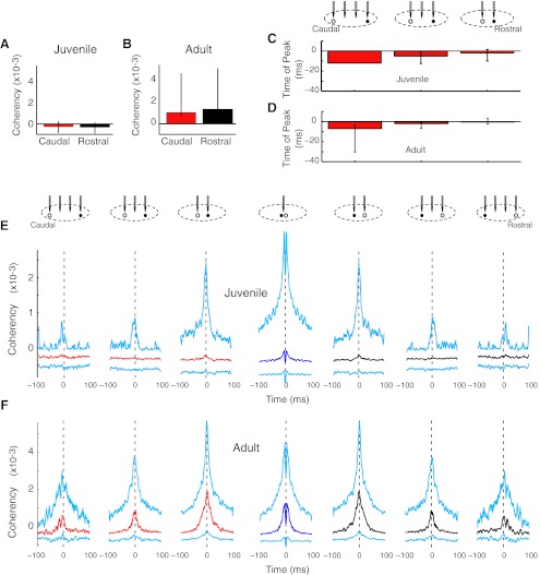
Principal neuron pairs did not show pronounced directionality in the strength of functional connectivity. A and B: neither juvenile nor adult principal neuron pairs had spatial differences in the magnitude of coherency. C and D: principal neuron pairs had rostrocaudal asymmetries in the time of peak coherency. In adults (D), this rostrocaudal pattern was significant. n = 1, 12, and 24 pairs for juvenilles (P = 0.43 by Kruskal-Wallis test); n = 3, 11, and 28 pairs for adults (*P < 0.05). E: median (caudal, red; same tetrode, dark blue; rostral, black) and IQR (cyan) coherencies are shown for juveniles. The caudal and rostral data are mirror images because coherency was computed for each principal neuron in the pair. n = 86, 194, 321, 410, 321, 194, and 86 pairs. F: in adults, principal neuron pairs did not show the strong rostrocaudal skew in magnitude characteristic of principal neurons versus the entire population of HVC neurons (see Fig. 4H). n = 32, 125, 184, 278, 184, 125, and 32 pairs.

