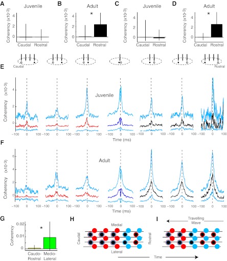
Fig. 7.
Principal neuron-interneuron functional connectivity was rostrocaudally skewed in adults but not in juveniles. A: the coherency of juvenile principal neurons with putative interneurons was not spatially distinct (n = 286 and 155 pairs). B: in contrast, adult principal neurons fired preferentially with rostral rather than caudal interneurons (n = 236 and 328 pairs, *P < 2e−6). C and D: a more restrictive definition of putative interneurons (<0.25-ms spike width for C and D only) yielded the same pattern of results in juveniles (n = 62 and 27 pairs) and adults (n = 35 and 221 pairs). E: in juveniles, no consistent pattern was observed in the spatial distribution of coherencies between principal neurons and putative interneurons. Median (caudal, red; same tetrode, dark blue; rostral, black) and IQR (cyan) coherencies are shown for juveniles. n = 37, 112, 137, 143, 107, 44, and 4 pairs. F: adult principal neurons were more functionally coactive with rostral interneurons than with those that were caudal to them. n = 31, 81, 124, 146, 148, 127, and 53 pairs. G: adult interneurons were more synchronized in the mediolateral axis than in the rostrocaudal axis. n = 105 and 12 pairs, *P < 0.038. H and I: one simple model that can explain the results posits a rostral-to-caudal travelling wave or advancing front of inhibition. In these images, the wave would travel right to left. The solid circles indicate inhibitory neurons. Principal neurons have a black center. Spiking activity is indicated by cyan and quiescence by red. Rostrocaudally oriented axons are indicated by black lines. Before the wave arrives, interneurons are relatively quiescent (red), whereas principal neurons may or may not be active (as indicated by red and cyan outlines). At the leading edge of the wave, all neurons are active. In the trailing wave, interneurons, but not principal neurons, are active.

