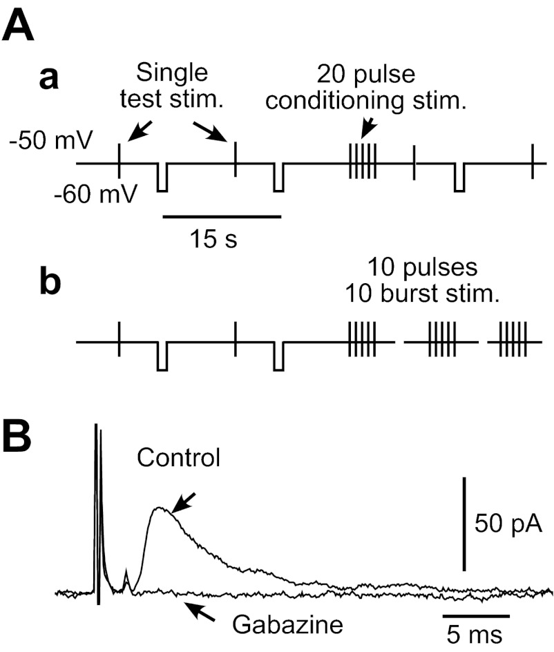
Fig. 1.
A: diagrams show stimulus (stim.) and the membrane-clamp schedules used in this study. Globus pallidus external segment (GPe) neurons were voltage clamped at −50 mV to block autonomous firing. Single test stimuli were given every 15 s, and −10 mV pulses were applied 5 s following each test stimulation to monitor the input impedance of the neuron. We used 2 repetitive stimulation schedules: single burst of 20 repetitive pulses with frequencies of 2–100 Hz (a) or 10 bursts of 10 repetitive pulses with 20 or 50 Hz and various interburst intervals (IBIs; b). B: stimulus intensity was adjusted to evoke 30–50 pA inhibitory postsynaptic currents (IPSCs) in test stimulation unless noted otherwise. For all recordings, artificial cerebrospinal fluid contained 10 μM 1,2,3,4-tetrahydro-6-nitro-2,3-dioxo-benzo[f]quinoxaline-7-sulfonamide and 30 μM 3-(2-carboxypiperzin-4-yl)-pro-pyl-1-phosponic acid to block AMPA/kainate and N-methyl-d-aspartate responses. Application of 10 μM gabazine completely abolished the fast IPSCs.

