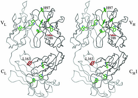
Fig. 1.
Stereoview of the crystal structure of 4C6 Fab, with the Cα trace of the light (L) and heavy (H) chains colored in light and dark gray, respectively. The modified tryptophan TrpL163 is highlighted in red, and other tryptophan residues (such as TrpH97) are colored green. The modified glutamine residue GlnH6 is also colored red. All of the figures were generated in bobscript (12) and rendered in raster3d (13).

