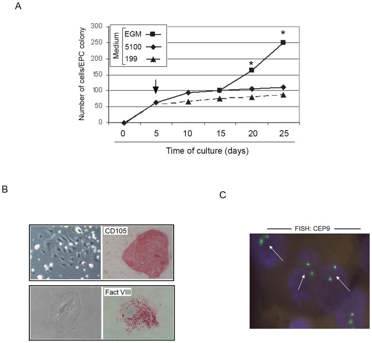
Figure 3. Identification of optimal culture conditions for the ex-vivo expansion of ACS PB-derived EPC/ECFC.
Primary EPC/ECFC colonies were generated by plating patient PBMC in M5100 medium, as detailed in the Method section. In A, after the colony identification (at day 5 after plating), medium was change (arrow) and replaced either with fresh M5100, or MEGM or M199 and the development of the colonies was monitored over the time. The growth kinetics of a representative experiment out of five independent experiments is shown. At each indicated time point, the mean cell number/ECFC was determined by two independent operators; standard deviations were below 10% and are not shown. Asterisk, p<0.05. In B, immunocytochemical analysis of in vitro expanded EPC/ECFC documenting positivity for CD105 antigen (original magnification: 20X) and for the specific endothelial marker Factor VIII (original magnification: 40X). In C, FISH analysis performed on in vitro expanded EPC/ECFC by using the centromeric enumeration probe CEP9 (white arrows) documenting a normal diploid chromosomal pattern (original magnification: 40X).

