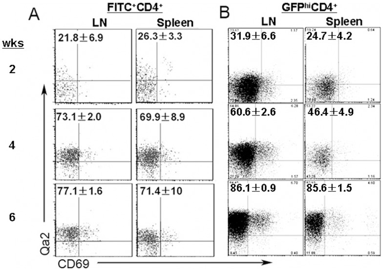
Figure 1. Pre-RTEs consist of SP4 thymocytes in adult mice but a mixture of SP3 and SP4 thymocytes in neonatal and young mice.
A. FITC intrathymic injection of C57BL/6 mice were used to analyze the phenotype of RTEs (FITC+ cells) at 2, 4, and 6 weeks of age. B. RAG2p-GFP mice were also used to study RTEs (GFPhi cells). The numbers in the plot represent the mean and standard deviation of the ratios of Qa2+CD69- cells in FITC+ or GFPhi CD4+CD8- T cells in the lymph nodes (LN) and spleen. Three to five mice were analyzed.

