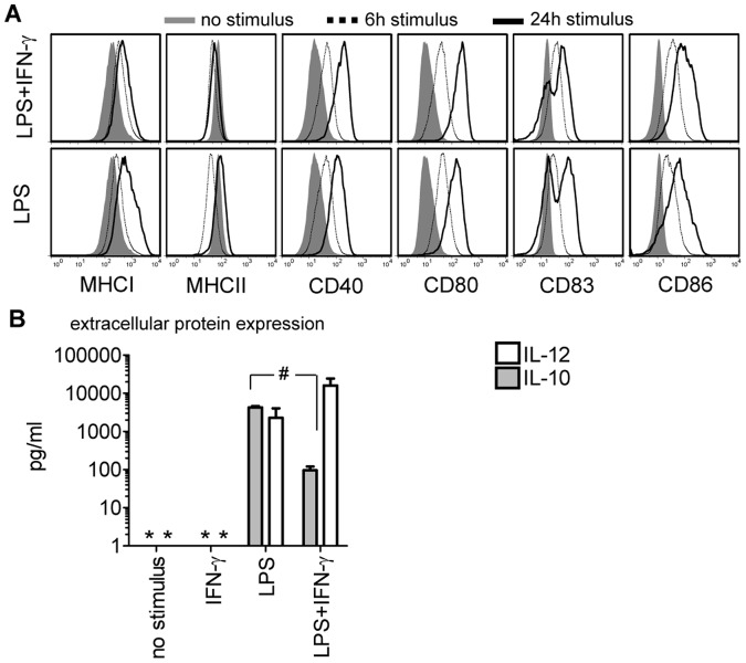
Figure 1. Detection of immune-modifying molecules in maturing human DCs.
(A) DC membrane molecule expression at 6 (dotted line) and 24 hours (black line) after LPS or LPS/IFN-γ exposure compared to un-stimulated human DCs (filled gray histograms). One representative out of 3 experiments is shown (donor C). (B) LPS or LPS/IFN-γ induced IL-12 (white bars) or IL-10 (grey bars) protein secretion was measured 48 hours after exposure to the different stimuli (p = 0.006). As a control, DCs were stimulated with IFN-γ or cultured without stimulation for 48 hours. Data are pooled from 4 experiments and depicted as median±SEM (donors A–D). Asterisks indicate cytokine levels below the detection limit of the assay.

