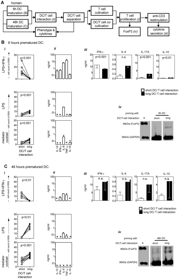
Figure 2. Proliferative capacity and polarization profile in short- versus long-term DC/T-cell co-cultures.
(A) Human DCs were matured for 6 or 48 hours, contacted with naïve allogeneic CD4+ enriched T-cells for 6 hours, separated using flow sorting, and either maintained alone in culture or re-cultivated with DCs (further-on referred to as “short”-term or “long”-term DC/T-cell interaction, respectively), both for 6 days. After the primary short- or long-term DC/T-cell co-cultures, T-cells were analyzed for FoxP3 expression or, adjusted to equal cell numbers, were re-stimulated with anti-CD3 for 24 hours. The supernatant of the re-stimulated culture was further analyzed for polarization-specific cytokine release. DCs matured for 6 (B) or 48 hours (C) with LPS, LPS/IFN-γ, or mediator cocktail (TNF-α/PG-E2/IL-1β/IL-6) were put in short-term (white squares) or long-term (black squares) co-culture with CD4+ T-cells. (i) Absolute numbers of proliferating CD4+CFSE−T-cells after 6 days of DC/T-cell co-culture are given. Pooled data from 8 to 10 experiments are shown. (ii) The cytokine composition in the supernatant of the 6 hours T-cell priming co-cultures with 6 or 48 hours matured DCs before sorting are shown. Asterisks indicate cytokine levels below the detection limit of the assay. Pooled data from 3 to 5 experiments are shown as median±SEM. Asterisks indicate cytokine levels below the detection limit of the assay. (iii) The culture supernatants of anti-CD3 re-stimulated CD4+ T-cells from short- (white squares) or long-term (black squares) primary DC/T-cell co-cultures with LPS/IFN-γ matured DCs (either 6 or 48 hours matured) were analyzed for their content of IFN-γ, IL-4, IL-17A, and IL-10. Three to 10 independent experiments are shown. (iv) Detection of FoxP3 in T-cells after short- or long-term DC/T-cell co-culture with LPS/IFN-γ stimulated DCs (matured for 6 or 48 hours). As a positive control (+) the cell lysate of a FoxP3 transfected cell line was used. One representative out of 3 experiments is shown. All data is given as median±SEM. DCs from Donors C, E and F were used in experiments (A) to (C).

