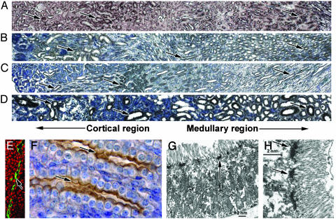
Fig. 3.
PKHD1 expression in the kidney. (A) Mouse adult kidneys were labeled by the same cRNA probe we used previously (12). Positive staining was observed in both the cortex and medulla regions (arrows, A). Corresponding regions of the kidney also showed positive staining (arrows) when the hAR-Np was used in mice (B), rats (C), and humans (D). Confocal microscopy using the hAR-Np reveals PKHD1 in an apical expression pattern in renal tubules (arrow, E); ethidium bromide (Sigma) was used for nuclei staining (red). The same pattern was observed in renal collecting ducts by using immunohistochemistry (arrows, F), but weak staining was also seen on the basolateral membrane. Electron microscopy using hAR-Np labeled with ImmunoGold indicates the expression of PKHD1 is localized to the terminal web below the microvilli along proximal tubule epithelial cells (arrows, G and H) (width, 5 μm, E; 10 μm, F; 150 μm, A–D).

