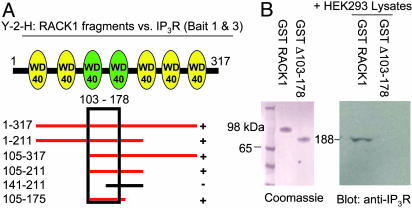
Fig. 2.
RACK1 binds to IP3R within WD40 repeats 3 and 4. (A) Cartoon of RACK1 depicting the seven WD40 repeats within the protein and the yeast two-hybrid analysis (Y-2-H) mapping of RACK1 to baits 1–345 and 346–922 of the IP3R. Red bars indicate positive binding to both IP3R fragments. (B Left) Coomassie stain of GST-purified RACK1 and mutant RACK1 Δ103–178. (Right) HEK293 cell lysates were passed over WT and Δ103–178 GST-RACK1 columns. Western analysis with anti-IP3R antibody demonstrates IP3R binding only to full-length RACK1.

