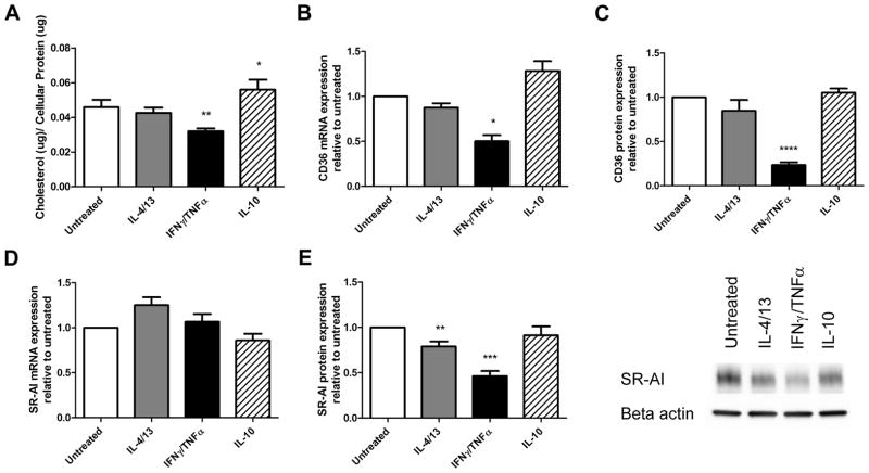
Figure 2. Macrophages treated with IFNγ and TNFα accumulate less cholesterol and are associated with a decrease in CD36 and MSR1 expression.
(A) Total cholesterol levels of different macrophage sub-phenotypes were measured after loading with 50ug/ml of oxLDL for 24 hours (n=6). mRNA levels of MSR1 (B), and CD36 (D) were measured by qRT-PCR and normalized to PPIB (n=4). Protein levels of MSR1 were determined by Western blotting (C) and shown with a representative blot (n=10). Cell surface CD36 protein expression was determined by flow cytometry (E), shown with a representative histogram (n=4). Results are expressed as means ± SEM of at least 3 different donors. Cholesterol accumulation was analyzed by one-way ANOVA followed by Tukey’s post-hoc test compared with untreated. mRNA and protein data was analyzed with a one-sample t-test. *p<0.05, **p<0.01, ***p<0.001.

