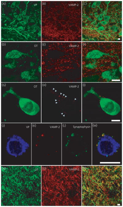
Fig. 1.

Vesicle associated membrane protein 2 (VAMP-2) immunoreactivity in supraoptic nucleus (SON) oxytocin (OT) and vasopressin (VP) neurones. Hypothalamic SON sections (A–F) and isolated neurones (G–M) show strong punctuate staining of VAMP-2 around VP and OT somata and dendrites; however, the overlays did not show co-localisation with the peptide. Labelling of VAMP-2 was seen on the outside of the plasma membrane of the neurones and co-localisation of VAMP-2 with synaptophysin (J–M) suggests labelling of pre-synaptic terminals. In the axon terminals in posterior pituitary (N–P), VAMP-2 is co-localised with the peptide, as shown by the yellow in the overlay (P). Arrowheads highlight examples of punctuate labeling around the outside an isolated cell. Scale bars = 10 μm.
