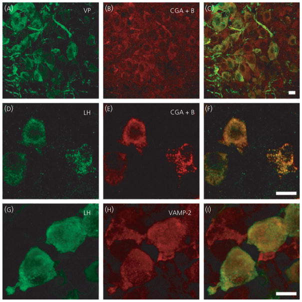
Fig. 2.
Positive controls for vesicle associated membrane protein 2 (VAMP-2) antibody and technique. Hypothalamic sections (A–C) show vasopressin (VP) and the large dense-cored vesicle protein chromogranin A (CGA)+B co-localised (C). In anterior pituitary sections (D–I), luteinising hormone (LH) is co-localised with both CGA+B and VAMP-2, as shown in the overlay images (F, I), demonstrating that the tissue and technique is not responsible for the absence of oxytocin and vasopressin co-localisation with VAMP-2 in cell somata and dendrites shown in previous figure. Scale bars = 10 μm.

