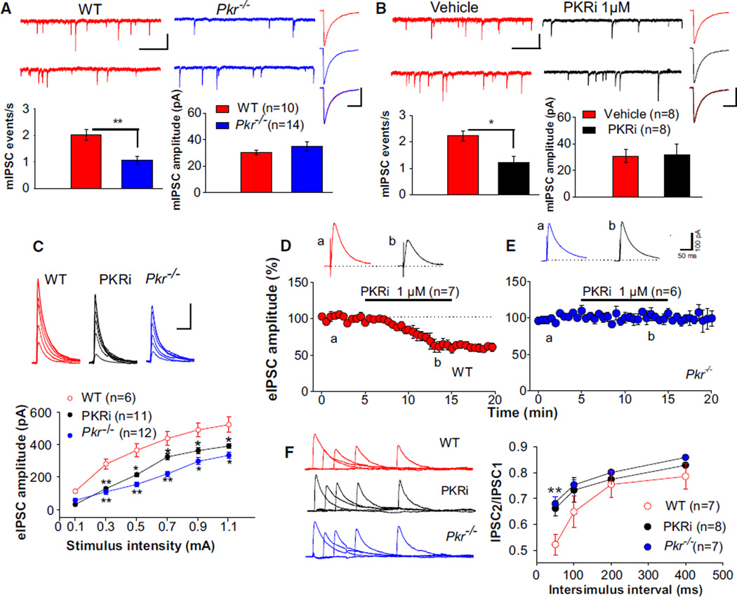
Figure 3. Reduced Inhibitory Synaptic Responses in CA1 of Pkr−/− Slices or WT Slices Treated with PKRi.
(A) Sample traces (top) and summary data (bottom) show reduced frequency (t = 4.7; **p < 0.01) but no change in the amplitude (Mann-Whitney U test, U = 66.0; p = 0.84) of mIPSCs (recorded at −60 mV with a KCl-containing patch pipette and in the presence of the wide-spectrum glutamate antagonist kynurenic acid [2 mM] and tetrodotoxin [TTX, 1 µM]) in CA1 neurons from Pkr−/− mice. Traces at right (each is an average of at least 100 mIPSCs) do not differ between WT (uppermost) and Pkr−/− slices (middle), as confirmed by superimposed traces (lowest).
(B) Similarly, in WT slices, PKRi decreased the frequency of mIPSCs (t = 3.42; *p < 0.05), but not their amplitude (t = 0.46; p = 0.65). Summary data and individual events are as in (A).
(A and B) Calibrations: 1 s and 50 pA for slow traces; 20 ms and 20 pA for fast traces.
(C) eIPSC amplitude as a function of stimulation intensity is shown superimposed and plotted (below) as input/output curves (recorded at 0 mV in the presence of APV [50 µM], CNQX [10 µM], and CGP55845 [10 µM]) (*p < 0.05; **p < 0.01). Calibrations: 50 ms and 200 pA.
(D and E) PKRi reduced the amplitude of evoked IPSCs in WT slices (D) (t = 3.2; p < 0.01), but not in Pkr−/− slices (E) (Mann-Whitney U test, U = 20.2; p = 0.62). Horizontal bar indicates PKRi application. Inset traces were obtained at times “a” and “b.”
(F) Paired IPSCs at 50, 100, 200, and 400 ms interstimulus intervals (ISIs) are superimposed (at left) after subtracting the first IPSC from paired responses. Corresponding ratios of IPSC2/IPSC1 are plotted at right. Note the reduced paired-pulse depression at 50 ms in Pkr−/− slices (t = 7.85; **p < 0.01) and WT slices treated with PKRi (t = 3.47; **p < 0.01).
See Figure S2 for further information about sIPSCs and eIPSCs in PKR-deficient slices; Figure S3 for the role of PKR in cumulative GABAergic inhibition; Figure S6A for the reduced mIPSCs in slices from eIF2α+/S51A mice; and Figure S7C for the effect of PKRi in vitro.

