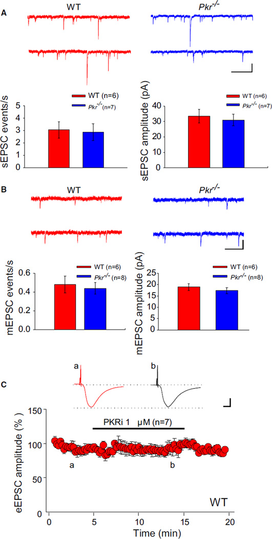
Figure 4. Excitatory Synaptic Transmission Is Unaltered in Pkr−/− Slices or WT Slices Treated with PKRi.
(A) Whole-cell recordings of EPSCs were performed with gluconate-containing pipettes at −70 mV in the presence of 100 µM picrotoxin. Sample traces (top) and summary data (bottom) show similar frequency (Mann-Whitney U test, U = 20.5; p = 0.95) and amplitude (t = 0.48; p = 0.64) of spontaneous EPSCs (sEPSCs) in WT and Pkr−/− slices.
(B) Sample traces (top) and summary data (bottom) show similar frequency (Mann-Whitney U test, U = 16.2; p = 0.34) and amplitude (t = 0.34; p = 0.74) of miniature EPSCs (mEPSCs) (recorded in the presence of picrotoxin [100 µM] and TTX [1 µM]) in WT and Pkr−/− slices.
(C) PKRi (1 µM) had no effect on EPSCs evoked (for “a” versus “b”; illustrated by inset traces, t = 0.40; p = 0.69) in the presence of 100 µM picrotoxin. Horizontal bar indicates the period of incubation with PKRi.
(A and B) Calibrations: 1 s and 20 pA. (C) Calibrations: 10 ms and 100 pA. For information about the effect of PKRi in vitro, see Figure S7C.

