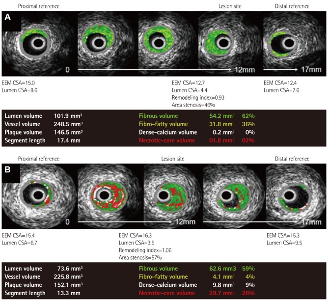
Fig. 1.
Representative VH-IVUS findings in both groups. A: normocholesterolemic patient (42 years old man with stable angina pectoris) showed small amounts of necrotic core volume. B: hypercholesterolemic patient (55 years old man with ST-segment elevation myocardial infarction) showed large amounts of necrotic core volume. VH-IVUS: virtual histology-intravascular ultrasound, EEM: external elastic membrane, CSA: cross sectional area.

